Human eye · मानव नेत्र
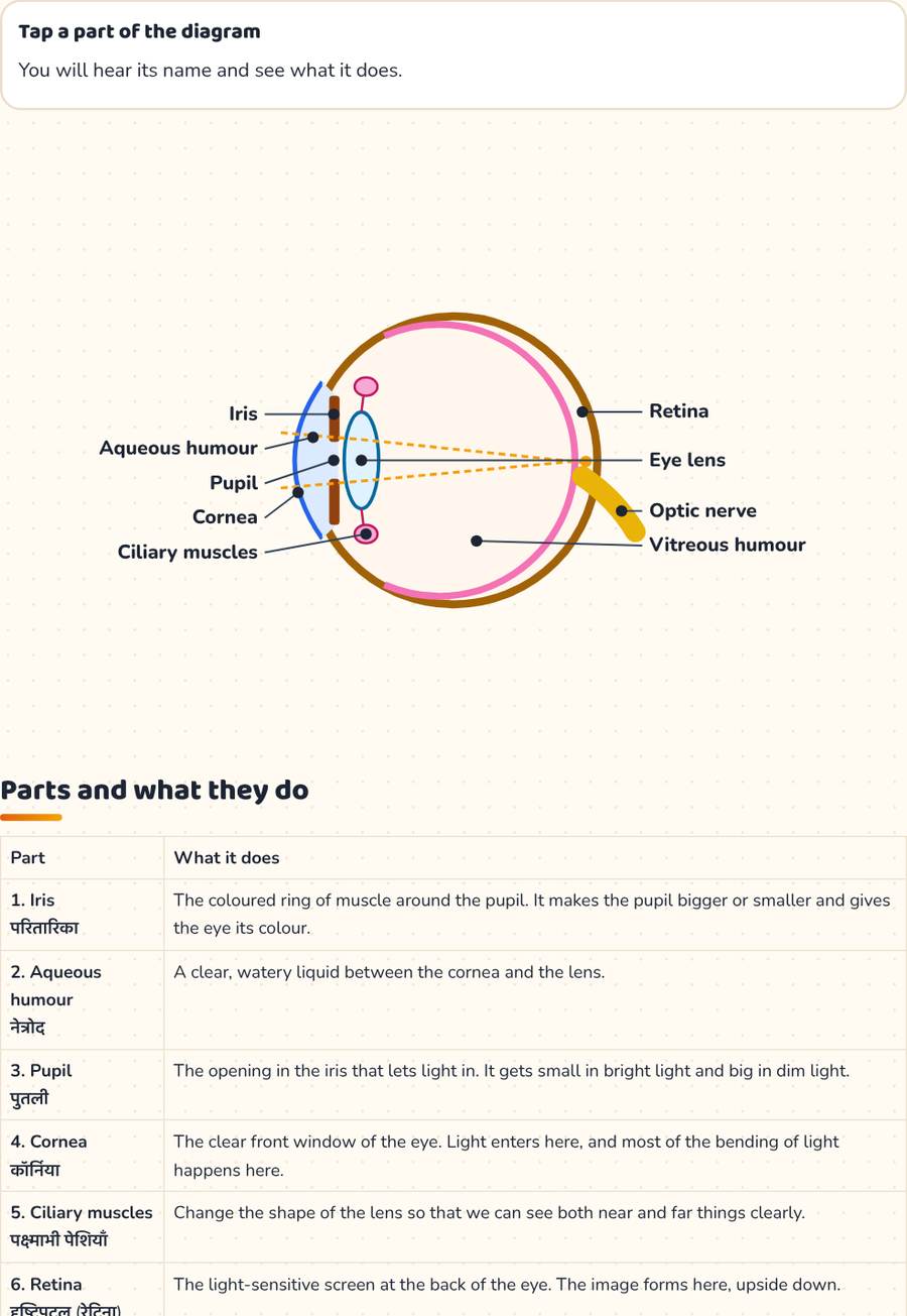
Our eyes let us see the world. Light enters through the clear cornea, passes through the pupil and the lens, and makes a picture on the retina. The optic nerve takes the message to the brain.
Human eye games
Tap a part of the diagram
You will hear its name and see what it does.
Parts and what they do
| Part | What it does |
|---|---|
| 1. Iris परितारिका | The coloured ring of muscle around the pupil. It makes the pupil bigger or smaller and gives the eye its colour. |
| 2. Aqueous humour नेत्रोद | A clear, watery liquid between the cornea and the lens. |
| 3. Pupil पुतली | The opening in the iris that lets light in. It gets small in bright light and big in dim light. |
| 4. Cornea कॉर्निया | The clear front window of the eye. Light enters here, and most of the bending of light happens here. |
| 5. Ciliary muscles पक्ष्माभी पेशियाँ | Change the shape of the lens so that we can see both near and far things clearly. |
| 6. Retina दृष्टिपटल (रेटिना) | The light-sensitive screen at the back of the eye. The image forms here, upside down. |
| 7. Eye lens अभिनेत्र लेंस | A clear, jelly-like lens that focuses light on to the retina. |
| 8. Optic nerve दृक् तंत्रिका | Carries messages from the retina to the brain, which makes sense of what we see. |
| 9. Vitreous humour काचाभ द्रव | A clear jelly that fills the eyeball behind the lens and keeps its round shape. |
The path of light
Cornea → Aqueous humour → Pupil → Eye lens → Vitreous humour → Retina → Optic nerve → Brain
Some facts
- The picture on the retina is upside down. The brain turns it the right way up.
- The retina has two kinds of cells: rods work in dim light and cones see colours.
- Where the optic nerve leaves the eye there are no light cells. This is the blind spot.
- Close one eye in a dark room and then in bright light: watch your pupil change size in a mirror.
Take care of your eyes
- Never look straight at the Sun.
- Read in good light, and take a break from screens every 20 minutes.
- Do not rub your eyes. Wash them with clean water if dust gets in.
The chart has the labelled diagram and what each part does. The worksheet has the diagram with blank labels and questions.
Human eye · मानव नेत्र
| Part | What it does |
|---|---|
| 1. Iris परितारिका | The coloured ring of muscle around the pupil. It makes the pupil bigger or smaller and gives the eye its colour. |
| 2. Aqueous humour नेत्रोद | A clear, watery liquid between the cornea and the lens. |
| 3. Pupil पुतली | The opening in the iris that lets light in. It gets small in bright light and big in dim light. |
| 4. Cornea कॉर्निया | The clear front window of the eye. Light enters here, and most of the bending of light happens here. |
| 5. Ciliary muscles पक्ष्माभी पेशियाँ | Change the shape of the lens so that we can see both near and far things clearly. |
| 6. Retina दृष्टिपटल (रेटिना) | The light-sensitive screen at the back of the eye. The image forms here, upside down. |
| 7. Eye lens अभिनेत्र लेंस | A clear, jelly-like lens that focuses light on to the retina. |
| 8. Optic nerve दृक् तंत्रिका | Carries messages from the retina to the brain, which makes sense of what we see. |
| 9. Vitreous humour काचाभ द्रव | A clear jelly that fills the eyeball behind the lens and keeps its round shape. |
kheloaurseekho.in/science/human-eye-diagram/
Human eye — worksheet
Name: ____________________ Date: __________
A. Label the parts of the diagram.
B. Answer these.
Answers: kheloaurseekho.in/science/human-eye-diagram/
More science: Human digestive system · Human respiratory system · Plant cell diagram · Animal cell diagram · Solar system · Photosynthesis · Human brain · Human heart · Parts of a flower · Parts of a plant · Periodic table · Water cycle